All posts by amaroneeditor

ESG 2015 (Prague)

ESG-2015Prague

ESG 2015 (Prague)

Long term management of uterine fibroids with Ulipristal Acetate
11th Congress of European Society of Gynecology. 21-24 October 2015.

Chairperson/s:

  • Genazzani Andrea R. (IT), Kucera Eduard (CZ)
  • Chabbert-Buffet Nathalie (FR) MoA and application of the SPRMs
  • Matule Dace (LV) PEARL IV Study: Latest results from the long term intermittent use of Ulipristal Acetate
  • Arriagada Pablo (CH) PEARL Extension II Study: Cumulative experience after 8 treatment courses with Ulipristal Acetate
  • Deans Anne (GB) Long Term management of UF with Ulipristal Acetate: Practical guidance for clinicians

Uterine Fibroids Diagnosis

Uterine fibroids are frequently diagnosed during pelvic examinations, and the size and location of the fibroids can usually be determined during a gynaecological examination, most commonly by ultrasound.

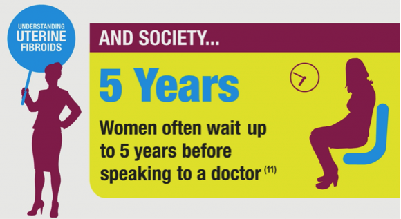
The time from the onset of symptoms to diagnosis can be up to five years19.

Uterine Fibroids Diagnosis

Uterine fibroids can be diagnosed in a number of different ways8 as transvaginal ultrasonography, magnetic resonance imaging, hysterosonography, hystersalpinography and hysteroscopy.

Understanding Uterine Fibroids

Uterine fibroids (uterine leiomyoma) are benign, monoclonal, hormone-sensitive, smooth muscle tumours of the uterus2, 8. They are the most common benign tumours of the female reproductive tract in pre-menopausal women.

While literature-reported incidence rates for uterine fibroids vary, in part due to the large proportion of women who go undiagnosed, uterine fibroids are estimated to affect between 20% and 40% of women of reproductive age2. Uterine fibroids are reported to be more frequent in the mature age group (40% to 70% of women between 35 years and 50 years) 9,10.

Although a number of risk factors have been linked to the development of uterine fibroids, the causes of uterine fibroids are not fully understood11. Factors such as African or Black-American ethnicity, older age, family history of uterine fibroids, nulliparity (the condition of never having given birth) or lower parity (number of times a woman has given birth), and obesity have all been associated with a higher risk of developing uterine fibroids 11, 12.

Uterine fibroids are often asymptomatic, but when symptomatic, the primary symptoms are heavy (uterine) bleeding, anaemia, abdominal pressure, abdominal pain, increased urinary frequency and infertility2. In particular, heavy menstrual blood loss is one of the most frequently disabling symptoms of uterine fibroids8.

Uterine Fibroids Economic Impact

Women with uterine fibroids experience significantly worse Health-Related Quality of Life (HRQoL) than women without fibroids3. Heavy menstrual bleeding (HMB) is common in women with symptomatic fibroids, and is associated with a considerable QoL burden, including psychological and social factors, and disruption to a normal routine39. Uterine fibroids impair productivity, increase absenteeism, and are associated with increased disability claims for symptomatic women40. In the CHASM study4 which was carried out in five European countries (France, Germany, Italy, Spain and UK) and used WPAI-SHP to measure absenteeism scores, employed women with uterine fibroids reported an absenteeism rate of 32.7%. Furthermore productivity was impaired by 36.1% and activity was impaired by 37.9%4.

Surgical treatment for uterine fibroids place a significant demand on health care resources. A further economic burden may be associated with the complications resulting from surgery, or the need for re-surgery, particularly in cases where the women have undergone UAE or myomectomy.

Hystersalpingography

Like hysterosonography, hysterosalpinography can be used to evaluate the extent of uterine fibroids. The procedure is carried out by use of a dye to highlight the uterine cavity and fallopian tubes in collaboration with X-ray. This procedure is mainly used where infertility is a problem, as a result of the fibroids, and this test can not only investigate the fibroids but also determine if the fallopian tubes are open or closed. The procedure like the hysterosonography is a relatively invasive procedure.

Hysterosonography

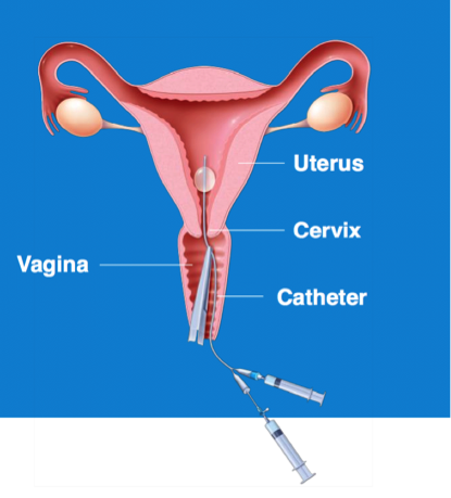
Hysterosonography, also known as a ‘saline infusion sonogram’, uses sterile saline to expand the uterine cavity, making it easier to obtain images of the uterine cavity and endometrium. This test is a useful tool when the subject is suffering from heavy menstrual bleeding despite normal ultrasound results. This procedure is also used to evaluate the extent of submucosal fibroids, but is a relatively invasive procedure.

Hysterosonography