Category Archives: Fibroids

Pharmacological Therapies

Approach Appropriate population Description
Esmya®
(Selective Progesterone Receptor Modulator, SPRM)
EU Market only
Young or premenopausal women
  • Orally administered
  • Treats multifactorial symptoms of uterine fibroids such as bleeding, amenorrhea, pain and fibroids size
  • Rapid and sustained reduction in heavy bleeding
  • Continuous reduction of fibroid size
  • Repeated 3-months treatment courses that need to be separated by off-drug interval
GnRH1 agonists Preoperative therapy in young or premenopausal women
  • Temporary treatment (3 to 6 months), fibroid re-growth on cessation
  • Flare effect during the first month
  • Route of administration: Injections
  • Adverse effects including loss of bone mineral density and menopausal symptoms
Pharmacological therapies used for treatment of Heavy Menstrual Bleeding (including LNG-IUS2, COC3, Oral progestogen, NSAIDs4 and tranexamic acid) Young or premenopausal women
  • Focused mainly on bleeding and /or pain. Limited or no effect on fibroid size
  • LNG-IUS are contra-indicated in women with distorted uterus
1 GnRH: Gonadotrophin releasing hormone

2 LNG-IUS: Levonorgesteral-releasing intrauterine system
3 COC: Combined oral contraceptive pill
4 NSAID: Non-steroidal anti-inflammatory drug

Uterine Fibroids Diagnosis

Uterine fibroids are frequently diagnosed during pelvic examinations, and the size and location of the fibroids can usually be determined during a gynaecological examination, most commonly by ultrasound.

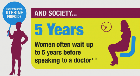
The time from the onset of symptoms to diagnosis can be up to five years19.

Uterine Fibroids Diagnosis

Uterine fibroids can be diagnosed in a number of different ways8 as transvaginal ultrasonography, magnetic resonance imaging, hysterosonography, hystersalpinography and hysteroscopy.

Understanding Uterine Fibroids

Uterine fibroids (uterine leiomyoma) are benign, monoclonal, hormone-sensitive, smooth muscle tumours of the uterus2, 8. They are the most common benign tumours of the female reproductive tract in pre-menopausal women.

While literature-reported incidence rates for uterine fibroids vary, in part due to the large proportion of women who go undiagnosed, uterine fibroids are estimated to affect between 20% and 40% of women of reproductive age2. Uterine fibroids are reported to be more frequent in the mature age group (40% to 70% of women between 35 years and 50 years) 9,10.

Although a number of risk factors have been linked to the development of uterine fibroids, the causes of uterine fibroids are not fully understood11. Factors such as African or Black-American ethnicity, older age, family history of uterine fibroids, nulliparity (the condition of never having given birth) or lower parity (number of times a woman has given birth), and obesity have all been associated with a higher risk of developing uterine fibroids 11, 12.

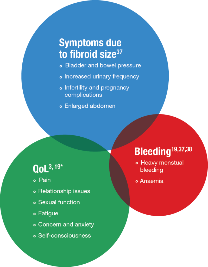
Uterine fibroids are often asymptomatic, but when symptomatic, the primary symptoms are heavy (uterine) bleeding, anaemia, abdominal pressure, abdominal pain, increased urinary frequency and infertility2. In particular, heavy menstrual blood loss is one of the most frequently disabling symptoms of uterine fibroids8.

Uterine Fibroids Economic Impact

Women with uterine fibroids experience significantly worse Health-Related Quality of Life (HRQoL) than women without fibroids3. Heavy menstrual bleeding (HMB) is common in women with symptomatic fibroids, and is associated with a considerable QoL burden, including psychological and social factors, and disruption to a normal routine39. Uterine fibroids impair productivity, increase absenteeism, and are associated with increased disability claims for symptomatic women40. In the CHASM study4 which was carried out in five European countries (France, Germany, Italy, Spain and UK) and used WPAI-SHP to measure absenteeism scores, employed women with uterine fibroids reported an absenteeism rate of 32.7%. Furthermore productivity was impaired by 36.1% and activity was impaired by 37.9%4.

Surgical treatment for uterine fibroids place a significant demand on health care resources. A further economic burden may be associated with the complications resulting from surgery, or the need for re-surgery, particularly in cases where the women have undergone UAE or myomectomy.

Hystersalpingography

Like hysterosonography, hysterosalpinography can be used to evaluate the extent of uterine fibroids. The procedure is carried out by use of a dye to highlight the uterine cavity and fallopian tubes in collaboration with X-ray. This procedure is mainly used where infertility is a problem, as a result of the fibroids, and this test can not only investigate the fibroids but also determine if the fallopian tubes are open or closed. The procedure like the hysterosonography is a relatively invasive procedure.

Hysterosonography

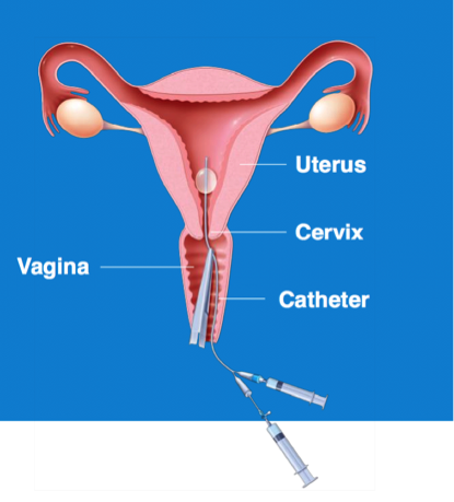
Hysterosonography, also known as a ‘saline infusion sonogram’, uses sterile saline to expand the uterine cavity, making it easier to obtain images of the uterine cavity and endometrium. This test is a useful tool when the subject is suffering from heavy menstrual bleeding despite normal ultrasound results. This procedure is also used to evaluate the extent of submucosal fibroids, but is a relatively invasive procedure.

Hysterosonography

Uterine Fibroid Symptoms

Click here to discover risks factors impacting uterine fibroids.

In most women symptoms are mild, and no treatment or pharmacological therapy will be required. However, some women experience symptoms that would be described as moderate or severe. Those women typically require pharmaceutical therapy.

Beyond their physical morbidity, uterine fibroids are a frequent cause of significant impairment of quality of life (QoL). In particular, abnormal uterine bleeding is one of the most frequently disabling symptoms of uterine fibroids, and this has associated physical, social and financial implications (including lower employment rates, more frequent absence from work, and loss of earnings in women). It is thought approximately 30% of symptomatic women suffer from abnormal bleeding which can negatively impact them due to feelings of self-consciousness and a reduction in their quality of life. Severe bleeding can also lead to severe iron deficiency, otherwise known as anaemia, which in its own right can cause fatigue, feelings of weakness and shortness of breath following exertion.

The main symptoms common with uterine fibroids are:


symptomsCommonWithUterineFibroids


* The Uterine Fibroid Symptom and Quality of Life (UFSQOL) questionnaire3,4.

Fibroids and fertility

Uterine fibroids, and in particular those which distort uterine anatomy and interfere with normal uterine physiology, can have adverse effect on reproductive function, leading to sub-fertility, later pregnancy complications, and miscarriage16. The incidence of fibroids in infertile women without any other obvious cause of infertility is estimated to be between 1% and 2.4%, however, there is a lack of prospective studies available to clearly determine the burden of fibroids in terms of their impact on fertility17.

A number of factors may influence fertility and pregnancy outcomes, including the size, location, and number of fibroids. Available evidence suggests that submucosal, intramural, and subserosal fibroids interfere with fertility in decreasing order of importance. Indeed, submucosal fibroids are linked to a 70% reduction in delivery rate17. There is some data suggesting an impact of the number and dimension of the lesions on fertility; in particular, fibroids over 5 cm in diameter, and located close to the cervix, are more likely to pose a problem16.

Classification of Uterine Fibroids

Uterine fibroids are traditionally classified according to their anatomical location as intramural, submucosal or subserosal. In addition, fibroids may be pedunculated (attached to the uterus by a stalk). Some larger fibroids may have characteristics of each type. Fibroids are not always isolated as there are often several coexistent lesions; in cases of multiple fibroids, a detailed ‘mapping’ of each lesion is valuable from a surgical point of view, although this may not always define a precise clinical situation14.

Fibroids can vary in size from millimetres to over 10 centimetres, and this can have a considerable clinical impact on the patient, with larger fibroids tending to respond less to pharmaceutical interventions, requiring more invasive surgical treatment options15.

Classification of Uterine Fibroids14, 16

  • Intramural fibroids: are fibroids which normally do not distort the uterine cavity and have <50% protruding into the serosal surface of the uterus and are the most common type. They typically develop within the wall of the uterus, beginning as small nodules in the muscular wall of the uterus. With time, intramural fibroids may expand inwards, causing distortion and elongation of the uterine cavity. They typically give the uterus a globular feeling on examination (like early pregnancy). They increase overall blood flow to the uterus and if large can distort and enlarge the internal cavity, even if they do not encroach onto it.
  • Subserosal fibroids: are those were >50% of the fibroid protrudes out of the serosal surface of the uterus. A subserosal fibroid may be sessile or pedunculated. They can grow quite large, but do not typically affect the size of the uterine cavity. They are more likely to produce pressure symptoms than heavy menstrual bleeding or infertility.
  • Submucosal fibroids: are defined as fibroids, which distort the uterine cavity according to the classification by the European Society of Hysteroscopy. The classification of these fibroids is then further divided into three subtypes: pedunculated fibroids without intramural extension (type 0), sessile (fixed in one location) with intramural extension of fibroid <50% (type I), and sessile with an intramural extension of >50% (type II). The degree of intramural extension can be assessed by ultrasonography or by hysteroscopy by observing the angle between the fibroid and the endometrium at the attachment to the uterine wall.

Submucosal fibroids are the fibroid type most likely to interfere with fertility14.

Other types of less frequent fibroids are:

  • Cervical fibroids: are found in the ‘neck’ (cervix) of the uterus and are difficult to remove without damaging the surrounding area.
  • Intraligamentous fibroids: are fibroids which may grow from a subserosal location into the peritoneal folds of the broad ligaments.